Parathyroid carcinoma with multiple brown tumors and pathological fracture: A case
report
Article / Artículo
https://doi.org/10.33821/780
Date received: 17/8/2025
Date accepted: 28/1/2026
Date: 01/4/2026
1. Introduction
The parathyroid glands consist of four glands that regulate serum calcium levels through
parathyroid hormone (PTH), which is produced by the chief cells. PTH acts on bones,
kidneys, and the intestine, promoting calcium release, reabsorption, and absorption,
respectively, as well as stimulating vitamin D production. Its secretion is regulated
by serum calcium and vitamin D levels through a negative feedback mechanism. Oxyphil
cells, which are present in smaller numbers, have no clearly defined endocrine function.
Hyperparathyroidism increases serum calcium levels and may be primary, secondary,
or tertiary, whereas hypoparathyroidism reduces calcium levels and is usually caused
by surgery. Both disorders severely affect mineral and neuromuscular balance [1].
Parathyroid carcinoma (PC) is a very rare endocrine malignancy, accounting for less
than 1% of cases of primary hyperparathyroidism and presenting with an extremely low
annual incidence. It can occur at any age, including in the pediatric population.
The mean age at diagnosis reported in most clinical series ranges from 51 to 57 years,
with no clear sex predilection. Its etiology is often unknown, although it has been
associated with genetic syndromes such as hyperparathyroidism-jaw tumor (HPT-JT) syndrome
and mutations in the CDC73 gene, among others [2,3].
Clinically, PC presents symptoms of severe hyperparathyroidism and may include a palpable
neck mass and serious metabolic complications. Definitive diagnosis requires histological
confirmation of tumor invasion, which makes preoperative identification challenging.
Early surgical intervention is the treatment of choice to improve prognosis. Due to
the rarity of this disease, case reports are valuable to expand clinical and therapeutic
knowledge [4].
This case report presents a clinical experience in Ecuador, it describes the diagnosis,
treatment, and outcome of a patient with parathyroid carcinoma. It aims at contributing
to the understanding and management of this rare disease in the local context.
2. Case report
A 27-year-old male patient with no relevant past medical or surgical history was referred
from a hospital within the Ecuadorian public health network due to persistent pain
in the right knee of approximately one year's duration, which worsened following a
traumatic event. After the trauma, radiographic studies of the affected limb revealed
an osteolytic lesion at the distal condyle of the right femur associated with a pathological
fracture. Based on the radiological characteristics of the lesion, osteosarcoma was
initially considered as presumptive diagnosis.
Upon hospital admission, the patient was conscious, hemodynamically stable, and reported
mild pain localized to the affected limb. Physical examination revealed grade 3 edema
according to the Godet scale and pain on deep palpation of the right knee. Initial
biochemical studies showed significant metabolic abnormalities, including serum calcium
of 16.9 mg/dL; ionized calcium, 7.94 mg/dL; creatinine, 1.22 mg/ dL; PTH level, 1604
pg/mL; and electrolyte disturbances such as potassium imbalance, hypomagnesemia, and
hypophosphatemia, as well as indirect hyperbilirubinemia. Given the clinical and biochemical
context, a whole-body computed tomography scan was performed, which revealed multiple
osteolytic lesions involving the left maxilla, distal condyle of the right femur with
pathological fracture, pelvis, scapulae, sternum, several thoracic vertebrae, and
bilateral costal arches, suggestive of disseminated bone disease.
A bone biopsy of the right femoral lesion was performed, and the histopathological
report revealed the presence of a brown tumor associated with hyperparathyroidism.
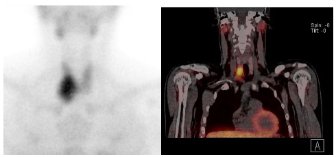
Based on these findings, parathyroid scintigraphy was requested, which demonstrated
an ectopic adenoma located in the right anterosuperior mediastinum (Figure 1).
Figure 1
Parathyroid scintigraphy: Hyperfunctioning ectopic parathyroid adenoma located in
the right anterosuperior mediastinum.
Source: SOLCA - Guayaquil
The patient underwent an exploratory cervicotomy followed by resection of a parathyroid
adenoma with intraoperative PTH monitoring according to the Miami criteria. The following
post-incision values were obtained: 1,529 pg/mL at 5 minutes; 297 pg/mL at 10 minutes;
and 231 pg/mL at 15 minutes. Intraoperative frozen section analysis was performed
and reported as negative for malignancy; macroscopic features were consistent with
benign pathology. However, subsequent immunohistochemical analysis demonstrated positivity
for CD31, thus confirming tumor vascular invasion and allowing reclassification of
the case as low-grade parathyroid carcinoma. Consequently, right hemithyroidectomy
was indicated as part of the oncologic surgical management.
Subsequently, due to the pathological lesion of the right femur, an orthopedic procedure
was performed. It included tumor curettage, bone cementation, and prophylactic osteosynthesis
of the affected limb. As part of the metabolic management, the patient received an
infusion of zoledronic acid. At present, the patient remains under multidisciplinary
outpatient follow-up and is receiving replacement therapy with calcitriol, calcium
carbonate, and cholecalciferol. Serum parathyroid hormone levels have remained within
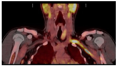
normal limits (PTH 58 pg/mL) with no clinical or biochemical evidence of disease recurrence
according to the most recent PET/CT report (Figure 2).
Figure 2
18F-choline PET/CT: Thyroidectomy. The left thyroid lobe shows a homogeneous appearance,
with no evidence of hypermetabolic foci suggestive of active functional lesions.
Source: SOLCA - Guayaquil
3. Discussion
Parathyroid carcinoma (PC) is a rare endocrine malignancy (<1% of primary hyperparathyroidism
cases) characterized by severe hypercalcemia (>17 mg/dL), markedly elevated PTH levels
(>1600 pg/mL), and significant skeletal involvement, including pathological fractures
and lytic lesions such as those observed in this patient [5,6].
Definitive diagnosis is established through histopathological examination of the complete
surgical specimen following parathyroidectomy. Although certain features-such as cytological
atypia or trabecular growth patterns-may suggest malignancy, the diagnosis requires
clear evidence of invasion into surrounding tissues or the presence of metastases.
According to the 2022 World Health Organization (WHO) classification, parathyroid
neoplasms are considered malignant when angioinvasion, lymphatic invasion, perineural
invasion, local invasion, or documented metastases are present [7]. Vascular invasion identified by CD31 is a known predictor of poor prognosis and
is associated with increased mortality and recurrence rates. Atypical parathyroid
tumors share some histological features with PC but lack definitive invasive characteristics.
Immunohistochemistry, particularly markers such as parafibromin, galectin-3, and a
Ki67 index >5%, supports the diagnostic process [8]. In cases where an initial simple resection is performed and malignancy is confirmed
postoperatively, surgical re-exploration with hemithyroidectomy is mandatory to ensure
tumor-free surgical margins [6,9].
These molecular and histopathological alterations not only contribute to understanding
tumor pathogenesis but also allow for individualized targeted therapies. Recent literature
highlights the emerging role of molecular biomarkers, such as circulating tumor cells,
as well as novel imaging modalities (including A18FFDG PET/CT and 4D CT) for advanced
staging and follow-up [10].
En bloc resection with negative margins remains the cornerstone of treatment, as simple
local excision is associated with high recurrence rates ranging from 8% to 51%. In
this case, complementary hemithyroidectomy was consistent with current evidence-based
recommendations [11,12].
Refractory hypercalcemia significantly contributes to morbidity and mortality. The
patient received zoledronic acid and calcium/vitamin D supplementation, in accordance
with current clinical guidelines [13]. In advanced cases, agents such as Cinacalcet, Denosumab, and even targeted therapies
have been evaluated in recent reports, showing favorable effects on calcium control
and tumor stabilization [14,15].
In patients with advanced or metastatic disease, promising responses have been reported
with tyrosine kinase inhibitors (TKIs) such as Sorafenib, Lenvatinib, Everolimus,
and Sunitinib, as well as combinations with Denosumab and calcimimetics-particularly
in patients harboring activating mutations in pathways such as PI3K/AKT/mTOR, KDM5C,
and CDC73 [16]. Although clinical trials remain limited, these therapies offer new perspectives,
especially in multidisciplinary referral centers [15].
Five-year survival rates range from 60% to 93%, largely dependent on effective hypercalcemia
control and complete surgical resection. The presence of bone metastases, as seen
in this case, requires a multidisciplinary surgical approach, including orthopedic
intervention, curettage, and cementation-to relieve symptoms and normalize calcium
levels, findings that are consistent with previously reported series [17]. Strict long-term follow-up with monitoring of serum calcium, PTH levels, and functional
and structural imaging is essential due to the high risk of late recurrence, ideally
in experienced centers with adequate endocrinologic and orthopedic support [18].
4. Conclusion
Parathyroid carcinoma is an uncommon but potentially lifethreatening endocrine neoplasm
characterized by severe hypercalcemia and serious skeletal complications. Its diagnosis
requires specific histological criteria, such as vascular permeation and capsular
invasion. En bloc surgical resection with negative margins remains the treatment of
choice. In advanced cases, adjuvant and targeted medical therapies have been incorporated
with encouraging results. A multidisciplinary approach is essential to control hypercalcemia,
stabilize bone lesions, and reduce recurrence. Survival depends largely on metabolic
control and complete tumor resection. Longterm followup is mandatory due to the high
rate of late recurrence.
1. Introducción
La paratiroides está compuesta por cuatro glándulas que regulan el calcio sérico mediante
la hormona paratiroidea (PTH), producida por las células principales. La PTH actúa
en huesos, riñones e intestino, promoviendo la liberación, reabsorción y absorción
de calcio, respectivamente, y estimulando la producción de vitamina D. Su liberación
está regulada por los niveles de calcio y vitamina D mediante retroalimentación negativa.
Las células oxífilas, presentes en menor cantidad, no tienen función endocrina clara.
El hiperparatiroidismo eleva el calcio sérico y puede ser primario, secundario o terciario,
mientras que el hipoparatiroidismo reduce el calcio y suele ser causado por cirugía.
Ambos trastornos afectan gravemente el equilibrio mineral y neuromuscular [1].
El carcinoma paratiroideo (CP) es una neoplasia endocrina rara, que representa menos
del 1% de los casos de hiperparatiroidismo primario y tiene una incidencia anual baja.
Se presenta a cualquier edad, incluida la población pediátrica. La edad media al momento
del diagnóstico es de 51-57 años en la mayoría de las series clínicas, sin preferencia
de sexo, y su causa suele ser desconocida, aunque está asociado con síndromes genéticos
como el de hiperparatiroidismo-tumor mandibular (HPT-JT) y mutaciones en el gen CDC73,
entre otros [2,3].
Clínicamente, el CP se manifiesta con síntomas de hiperparatiroidismo severo y puede
incluir masa palpable y complicaciones metabólicas graves. El diagnóstico definitivo
requiere confirmación histológica de invasión tumoral, lo que dificulta su identificación
preoperatoria. La cirugía temprana es el tratamiento de elección para mejorar el pronóstico,
y debido a la rareza de esta enfermedad, reportes de casos son importantes para ampliar
el conocimiento clínico y terapéutico [4].
Este reporte de caso presenta una experiencia clínica en Ecuador, en él se describe
el diagnóstico, el tratamiento y la evolución de un paciente con carcinoma paratiroideo,
con el objetivo de contribuir al conocimiento y manejo de esta rara enfermedad en
el contexto local.
2. Caso clínico
Un paciente masculino de 27 años, sin antecedentes patológicos ni quirúrgicos de relevancia,
fue remitido de un hospital de la red de salud pública del Ecuador por cuadro de dolor
persistente en la rodilla derecha de aproximadamente un año de evolución, reagudizado
luego de un evento traumático. A raíz de este evento, se realizaron estudios radiográficos
de la extremidad, en los cuales se evidenció una lesión osteolítica en el cóndilo
femoral distal derecho, asociada a fractura patológica. Por las características radiológicas
de la lesión, se planteó como diagnóstico presuntivo un osteosarcoma.
A su ingreso hospitalario, el paciente se encontraba consciente, hemodinámicamente
estable y refería dolor leve localizado en la extremidad afectada. El examen físico
reveló edema grado 3 según escala de Godet y dolor a la palpación profunda en la rodilla
derecha. Los estudios bioquímicos iniciales mostraron alteraciones metabólicas relevantes:
calcio sérico 16,9 mg/dL, calcio iónico 7,94 mg/dL, creatinina 1,22 mg/ dL, PTH 1604
pg/mL y trastornos electrolíticos (potasio, hipomagnesemia e hipofosfatemia), además
de hiperbilirrubinemia indirecta. Dado el contexto clínico y bioquímico, se realizó
tomografía computarizada de cuerpo entero, la cual reportó múltiples lesiones osteolíticas,
localizadas en maxilar izquierdo, cóndilo distal del fémur derecho con fractura patológica,
pelvis, escápulas, esternón, varias vértebras dorsales y arcos costales bilaterales,
sugestivas de enfermedad ósea diseminada.
Se realizó una biopsia ósea de la lesión en el fémur derecho, cuyo resultado histopatológico
informó la presencia de un tumor pardo, hallazgo relacionado con hiperparatiroidismo.
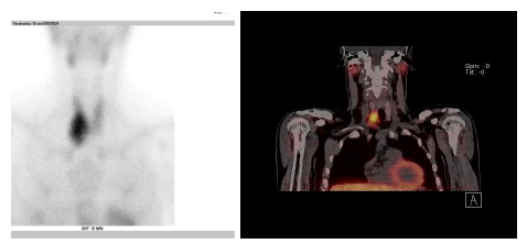
Ante estos hallazgos se solicitó gammagrafía paratiroidea, la cual evidenció un adenoma
ectópico ubicado en mediastino anterosuperior derecho (Figura 1).
Figura 1
Gammagrafía paratiroidea: adenoma paratiroideo ectópico hiperfuncionante en el mediastino
anterosuperior derecho.
Fuente: SOLCA - Guayaquil
El paciente fue sometido a cervicotomía exploradora más resección de adenoma paratiroideo
con protocolo PTH intraoperatoria mediante criterios Miami. Se obtuvieron los siguientes
valores posincisión: 1529 pg/mL a los 5 minutos; 297 pg/mL a los 10 minutos, y 231
pg/mL a los 15 minutos. También se obtuvo biopsia por congelación negativa para malignidad
con características macroscópicas compatibles con patología benigna. No obstante,
el estudio de inmunohistoquímica posterior evidenció positividad para CD31, que confirmó
permeación vascular tumoral y permitió establecer el diagnóstico de carcinoma de paratiroides
de bajo grado de diferenciación. En consecuencia, se realizó hemitiroidectomía derecha
como parte del tratamiento quirúrgico oncológico.
Luego, debido a la lesión patológica en el fémur derecho, se realizó procedimiento
ortopédico consistente en vaciamiento tumoral, cementación ósea y osteosíntesis profiláctica
de la extremidad. Como parte del manejo metabólico, recibió infusión de ácido zoledrónico.
Actualmente, el paciente continúa en seguimiento ambulatorio multidisciplinario, bajo
tratamiento de reemplazo con calcitriol, carbonato de calcio e hidroferol; mantiene
cifras séricas de hormona paratiroidea (PTH 58 pg/mL) dentro de parámetros normales
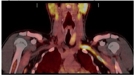
y sin evidencia clínica ni bioquímica de recurrencia de la enfermedad según el último
reporte de PET/CT (Figura 2).
Figura 2
PET CT con 18F-colina: tiroidectomía derecha. Se observa lóbulo tiroideo izquierdo
de aspecto homogéneo, sin evidencia de focos hipercaptantes que sugieran lesiones
funcionales activas.
Fuente: SOLCA - Guayaquil
3. Discusión
El CP es una neoplasia endocrina rara (< 1 % de hiperparatiroidismo primario) que
se caracteriza por hipercalcemia grave (≥ 17 mg/dL), PTH marcadamente elevada (> 1600
pg/mL) y afectación ósea franca; incluye también fracturas patológicas y lesiones
líticas como las encontradas en este paciente [5,6].
El diagnóstico definitivo se realizó mediante el examen histopatológico de la pieza
completa tras la paratiroidectomía. Aunque algunas características, como atipia citológica
o crecimiento trabecular, sugieren malignidad, el diagnóstico requiere evidencia clara
de invasión a tejidos circundantes o metástasis. Según la Organización Mundial de
la Salud (2022), se consideran malignas las neoplasias con angioinvasión, invasión
linfática, perineural, local o metástasis documentadas [7]. La invasión vascular identificada por CD31 es un predictor de mal pronóstico, asociada
a mayor mortalidad y recurrencia. Los tumores atípicos comparten rasgos con el CP,
pero sin invasión evidente. La inmunohistoquímica, especialmente con parafibromina,
galectina-3 y Ki-67 > 5 %, apoya el diagnóstico[8]. En casos en los que inicialmente se realiza una resección simple y posteriormente
se confirma la malignidad, es bligaotoria una exploración quirúrgica con hemitiroidectomía
para asegurar márgenes quirúrgicos libres [6,9].
Estas alteraciones no solo ayudan en la patogénesis, sino que también permiten individualizar
terapias dirigidas. La literatura reciente destaca el uso emergente de biomarcadores
moleculares como células tumorales circulantes y nuevas modalidades de imagen (18F-FDG
PET/CT, 4DCT) para estadificación avanzada [10].
La resección en bloque con margen negativo sigue siendo el pilar del tratamiento,
ya que una escisión local simple conlleva altos índices de recidiva, del 8 % al 51
%. En este caso, la hemitiroidectomía complementaria se ajusta a las recomendaciones
basadas en evidencia [11,12].
La hipercalcemia refractaria contribuye significativamente a la morbimortalidad. Este
paciente recibió zoledronato y suplemento de calcio/vitamina D, lo que se alinea con
las guías actuales [13]. En casos avanzados, opciones derivadas como cinacalcet, denosumab o incluso terapia
dirigida se han estudiado en reportes recientes con efectos favorables en el control
del calcio y la estabilización tumoral [14,15].
En tumores avanzados o metastásicos, se reportan respuestas prometedoras de inhibidores
de la tirosina quinasa (TKIs) como sorafenib, lenvatinib, everolimus, sunitinib, y
combinaciones con denosumab y calcimiméticos, especialmente en pacientes con mutaciones
activadoras (PI3K/AKT/mTOR, KDM5C, CDC73) [16]. Aunque los ensayos clínicos aún son limitados, estas terapias ofrecen nuevas esperanzas,
sobre todo en centros de referencia multidisciplinarios [15].
La supervivencia a cinco años varía entre 60-93 %, condicionada por el control de
hipercalcemia y la resección completa. La presencia de metástasis óseas, como en este
caso, exige abordaje quirúrgico múltiple (ortopedia, vaciamiento, cementación), crucial
para aliviar síntomas y normalizar el calcio, lo que coincide con observaciones de
series revisadas [17]. Un seguimiento estricto en un centro con experiencia y adecuado soporte endocrinológico
y ortopédico, con monitoreo de calcio, PTH e imágenes funcionales y estructurales,
es esencial por la alta tasa de recidiva tardía [18].
4. Conclusión
El carcinoma de paratiroides es una neoplasia endocrina infrecuente pero potencialmente
letal, caracterizada por hipercalcemia severa y complicaciones óseas graves. Su diagnóstico
requiere criterios histológicos específicos, como permeación vascular e invasión capsular.
La resección quirúrgica en bloque con márgenes negativos sigue siendo el tratamiento
de elección. En casos avanzados, se han incorporado terapias médicas adyuvantes y
dirigidas con resultados alentadores. El manejo multidisciplinario es imprescindible
para controlar la hipercalcemia, estabilizar lesiones óseas y reducir recidivas. La
supervivencia depende del control metabólico y de una resección completa. Un seguimiento
prolongado es indispensable dada la alta tasa de recurrencia tardía.