Oncologic neurosurgery in the era of precision medicine and the challenges ahead
Article / Artículo
https://doi.org/10.33821/865
Date received: 02/1/2026
Date accepted: 31/3/2026
Date: 04/1/2026
Oncologic neurosurgery may be regarded as a medical subspecialty devoted to the diagnosis,
surgical treatment, and follow-up of primary and metastatic tumors of the central
nervous system (brain and spinal cord) in patients of all ages.
To fulfill this purpose, the oncologic neurosurgeon has to make objective use of neurosurgical
techniques and procedures adapted to oncology; that is, the diagnosis and treatment
of tumors involve a multidisciplinary team including neurosurgeons, neurologists,
oncologists, and radiation oncologists to address both cancer and its neurological
complications, using advanced imaging, genetics, and personalized therapy [1,2].
Advanced imaging techniques, artificial intelligence (AI), and molecular biology are
used to maximize tumor resection and preserve brain function. Technologies such as
neuronavigation, intraoperative imaging (MRI/ ultrasound), and functional mapping
(awake craniotomy) enable personalization, reduce sequelae, and improve survival [3-5].
Over time, through sustained and proactive effort, SOLCA-Guayaquil and its Neurosurgery
Service have made these advances available to patients. Human and technological resources
have also positioned them at the forefront in the country, while consistently striving
to maintain a level comparable to that of referral centers in other regions. All of
this with the main purpose of offering solutions for complex neuro-oncologic conditions
according to the particularities of each case.
There are several factors or subsystems that interact synergistically with technology
and knowledge, as well as with the results derived from their application over time,
and they are reflected in accumulated experience and effort. In this regard, significant
progress has been achieved, including the following subsystems [6-11]:
-
Neuronavigation and Augmented Reality: Brain GPS-type systems integrate preoperative
images with the real surgical field, thus enabling minimally invasive approaches.
-
Intraoperative Ultrasound: It complements the accuracy of the neuronavigation system.
It allows correction of the millimetric deviations that may occur with neuronavigation
during surgery and confirms that tumor resection has been achieved as planned (total
or partial).
-
Intraoperative Imaging and Navigation: The use of intraoperative MRI and high-resolution
ultrasound corrects the brain shift phenomenon (brain deformation after opening the
skull), thus enabling more complete resections.
-
Awake Craniotomy and Functional Mapping: This is used to identify eloquent brain areas
(language, movement) while the patient is awake, thus reducing the risk of neurological
deficits.
-
Molecular and Genomic Diagnosis: Real-time sequencing (for example, nanopore sequencing)
classifies tumors in less than one hour during surgery and helps determine the extent
of resection.
-
Fluorescence-Guided Surgery: Techniques such as the use of 5-ALA (5-aminolevulinic
acid) make tumor cells "glow" under specific lights and facilitates their visual identification
against healthy tissue.
-
PET Scan: Through radioactively labeled molecules such as glucose and citicoline,
PET imaging makes it possible to observe the metabolic behavior of tumors. The greater
their aggressiveness and growth, the greater their metabolic activity on the study.
Consequently, it helps obtain better biopsy samples in certain tumors for improved
identification-higher tumor grade or malignancy. It also allows staging or assessment
of tumor spread throughout the body in cases of brain metastases.
-
High-Precision Radiotherapy: Techniques such as radiosurgery, including robotic systems
and CyberKnife, or proton therapy provide high precision when delivering radiation,
thereby protecting healthy brain tissue.
-
Minimally invasive spine surgery: it substantially improves patients' quality of life.
At SOLCA-Guayaquil, resources aimed at achieving a high degree of precision are broadly
available; however, intraoperative magnetic resonance imaging is not available, although
in practice this limitation is reasonably offset by other previously mentioned tools
and modalities. Likewise, magnetic resonance imaging is performed within the first
24 postoperative hours to establish the degree of tumor resection. Subsequently, the
patient is prepared to receive adjuvant treatment with chemotherapy, radiotherapy,
or both, depending on each case.
In this context, oncologic neurosurgery (Figure 1) in the era of precision medicine has evolved from a predominantly extractive approach
to a model centered on molecular biology and the use of innovative technologies, aimed
at maximizing tumor resection without compromising neurological functions.
Figure 1
Technology in oncologic neurosurgery to achieve results safely and effectively.
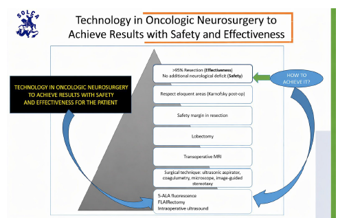
FLAIR: Fluid-Attenuated Inversion Recovery. FLAIRectomy or resection of the peritumoral
FLAIR hyperintensity An important aspect to be considered, in addition to precision,
is safety and effectiveness.
Safety is a fundamental concept because it makes it possible to minimize, to the greatest
extent possible, the side effects or collateral effects derived from any intervention.
In the context of oncologic neurosurgery, this implies avoiding the appearance of
new neurological deficits. More importantly, the higher objective is not only to preserve
neurological function, but also to improve preexisting symptoms and deficits that
compromise the patient's quality of life through a precise, planned intervention supported
by the best available evidence [12,13].
What lies ahead is fully personalized oncologic neurosurgery and molecular biology.
This era of precision will be achieved through the genomic analysis of each patient,
for instance:
-
Molecular Profiling: Next-Generation Sequencing (NGS) identifies specific mutations
to guide targeted therapies and predict treatment response.
-
Liquid Biopsy: It enables monitoring tumor dynamics through the analysis of DNA in
blood or cerebrospinal fluid in a minimally invasive manner.
-
Artificial Intelligence: AI algorithms assist in rapid diagnosis and real-time tumor
classification during surgery, supported by big data, Single-Cell Spatial Multiomics,
and molecular imaging.
In this transformation scenario, oncologic neurosurgery is moving toward an increasingly
precise, safe, and personalized model supported by the integration of molecular biology,
artificial intelligence, and innovative technologies. These open a more hopeful perspective
for patients with tumors of the central nervous system by translating into better
outcomes and quality of life.
La neurocirugía oncológica podría considerarse una subespecialidad médica dedicada
al diagnóstico, tratamiento quirúrgico y seguimiento de tumores primarios y metastásicos
del sistema nervioso central (cerebro y médula espinal) en pacientes de todas las
edades.
El neurocirujano oncológico requiere, para llevar a efecto este propósito, usar de
forma objetiva técnicas neuroquirúrgicas y procedimientos que se adecuan al campo
de la oncología; es decir, en el diagnóstico y tratamiento de los tumores participa
un equipo multidisciplinario que involucra a neurocirujanos, neurólogos, oncólogos
y radioterapeutas para abordar tanto el cáncer como sus complicaciones neurológicas,
utilizando técnicas avanzadas de imagen, genética y terapia personalizada [1,2].
Las técnicas avanzadas de imagen, inteligencia artificial (IA) y biología molecular
se utilizan para maximizar la resección tumoral y preservar funciones cerebrales.
Tecnologías como la neuronavegación, la imagenología intraoperatoria (MRI/ecografía)
y el mapeo funcional (craneotomía con el paciente despierto) permiten personalización,
lo que reduce secuelas y mejora la supervivencia [3-5].
El Hospital SOLCA-Guayaquil y el Servicio de Neurocirugía han logrado, a través del
tiempo y mediante un esfuerzo propositivo sostenido, poner a disposición de los pacientes
estos avances. Asimismo, cuentan con recursos humanos y tecnológicos que les han permitido
posicionarse a la vanguardia en el país, procurando de manera constante mantener un
nivel equiparable al de centros de referencia en otras latitudes. Todo ello con el
firme propósito de ofrecer la resolución de complejos padecimientos neurooncológicos,
según las particularidades de cada caso.
Existen diversos factores o subsistemas que interactúan de manera sinérgica con la
tecnología y el conocimiento, así como con los resultados que se derivan de su aplicación
a lo largo del tiempo, reflejados en la experiencia y el esfuerzo acumulados. En este
sentido, se han logrado avances significativos en este proceso. Estos subsistemas
incluyen [6-11]:
-
Neuronavegación y realidad aumentada: Sistemas tipo «GPS cerebral», integran imágenes
preoperatorias en el campo quirúrgico real que permiten abordajes de mínima invasión.
-
Ultrasonido intraoperatorio: Complementa la certeza del sistema neuronavegador. Permite
corregir las desviaciones de milímetros que pueden ocurrir con el neuronavegador durante
la cirugía y confirma si hay resección tumoral de acuerdo con lo planificado (total
o parcial).
-
Imagenología intraoperatoria y navegación: El uso de RM intraoperatoria y ecografía
de alta resolución corrige el fenómeno de brain shift (deformación cerebral tras abrir
el cráneo), y permite resecciones más completas.
-
Craneotomía con paciente despierto y mapeo funcional: Se utiliza para identificar
áreas cerebrales elocuentes (lenguaje, movimiento) mientras el paciente está despierto.
Esto reduce el riesgo de déficits neurológicos.
-
Diagnóstico molecular y molecular/genómico: La secuenciación en tiempo real (por ejemplo,
nanoporos) clasifica tumores en menos de una hora durante la cirugía, y esto ayuda
a determinar la extensión de la resección.
-
Cirugía guiada por fluorescencia: Técnicas como el uso de 5-ALA (ácido 5-aminolevulínico)
hacen que las células tumorales «brillen» bajo luces específicas, facilitando su identificación
visual frente al tejido sano.
-
PET Scan: Permite a través de moléculas radioactivamente marcadas como glucosa y citicolina
observar el comportamiento metabólico de los tumores. A mayor agresividad y crecimiento
tienen más actividad metabólica en el estudio. Como consecuencia de lo anterior, permite
tomar mejores muestras de biopsia en ciertos tumores para una mejor identificación
(mayor grado o malignidad del tumor). Además, permite el estadiaje o extensión del
tumor en el cuerpo en caso de metástasis cerebrales.
-
Radioterapia de alta precisión. Técnicas como la radiocirugía, incluidas la robótica
y la CyberKnife, o la terapia de protones ofrecen alta precisión al administrar radiación,
protegiendo el tejido cerebral sano.
-
Cirugía de columna mínimamente invasiva: que mejora sustancialmente la calidad de
vida de los pacientes.
En el Hospital SOLCA-Guayaquil se cuenta con una amplia disponibilidad de recursos
orientados a alcanzar un alto grado de precisión; sin embargo, no se dispone de resonancia
magnética intraoperatoria, la cual es, en la práctica, razonablemente suplida mediante
otras herramientas y modalidades previamente ya mencionadas. Asimismo, se realiza
resonancia magnética dentro de las primeras 24 horas del posoperatorio con el fin
de establecer el grado de resección tumoral. Posteriormente, el paciente es preparado
para recibir tratamiento coadyuvante con quimioterapia o radioterapia o ambos tratamientos
según cada caso.
En este contexto, la neurocirugía oncológica (Figura 1) en la era de la medicina de precisión ha evolucionado desde un enfoque predominantemente
extractivo hacia un modelo centrado en la biología molecular y en el uso de tecnologías
de vanguardia, orientado a maximizar la resección tumoral sin comprometer la funcionalidad
neurológica.
Un aspecto importante para considerar además de la precisión es la seguridad y efectividad.
Figura 1
Tecnología en neurocirugía oncológica.
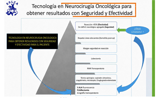
FLAIR: Fluid-Attenuated Inversion Recovery. FLAIRectomía o resección de la hiperintensidad
peritumoral en FLAIR.
La seguridad es un concepto fundamental, ya que permite minimizar, en la medida de
lo posible, los efectos secundarios o colaterales derivados de cualquier intervención.
En el contexto de la neurocirugía oncológica, esto implica evitar la aparición de
nuevos déficits neurológicos. Más aún, el objetivo superior es no solo preservar la
función neurológica, sino también mejorar los síntomas y déficits preexistentes que
comprometen la calidad de vida del paciente, mediante una intervención precisa, planificada
y sustentada en la mejor evidencia disponible [12,13].
Entre las tendencias futuras está la neurocirugía oncológica totalmente personalizada
y la biología molecular. Esta era de precisión se conseguirá por el análisis genómico
de cada paciente:
-
Perfilado molecular: La secuenciación de nueva generación (NGS) identifica mutaciones
específicas para guiar terapias dirigidas y predecir la respuesta al tratamiento.
-
Biopsia líquida: Permite monitorizar la dinámica tumoral mediante el análisis de ADN
en sangre o líquido cefalorraquídeo de forma mínimamente invasiva.
-
Inteligencia artificial: Algoritmos de IA asisten en el diagnóstico rápido y la clasificación
de tumores en tiempo real durante la cirugía (big data, single-cell spatial multiomics,
molecular imaging).
En este escenario de transformación, la neurocirugía oncológica avanza hacia un modelo
cada vez más preciso, seguro y personalizado, sustentado en la integración de la biología
molecular, la inteligencia artificial y las tecnologías de vanguardia. Esto abre una
perspectiva con mejores desenlaces clínicos para los pacientes con tumores del sistema
nervioso central, al traducirse en mejores resultados y calidad de vida.